Brain Imaging - Diffusion MRI

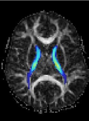
Magnetic Resonance Diffusion Tensor Imaging (MR-DTI) is a relatively new MR imaging technique which is based on measuring the extend of diffusing water molecules in 3D space. The technique is solely based on measuring the phase differences (via signal suppression due to phase differences) between spinning water molecules. This phase difference occurs due to the diffusion process because different molecules diffusing in different directions experience different magnetic fields (due to the spatially varying B-field). The output is a set of Diffusion Weighted Images (DWI). The diffusion tensor is mathematical structure that summarizes (upto second order accuracy) this diffusion process, i.e. the information in this set of diffusion weighted images. We suggest R. Bammer's, from Stanford University LUCAS MRS/I Center, PhD thesis as a complete reference for MR-DTI. A fundamental problem related to DTI is that you need to monitor (listen to the MR signal) a sufficiently large volume (voxel), after waiting for sufficiently long time to see the effect of anisotropic diffusion (if there is any). Furthermore DTI itself is only a 2nd order approximate modeling of the true diffusion process. These issues impose limitations of the speed and resolution of DTI. In other words, DTI can not and will not be able to image single fibers. Consequently, the interpretation of DTI data needs reliable modeling regarding the information it contains.

DTI based research at VAVlab is focused on the analysis and visualization of 3D DTI tensor fields as acquired by coventional and commercially available MRI systems. The subtopics that we work on are (i) tensor and vector field regularization, (ii) fiber tractography and volume connectivity analysis, (iii) interactive user interfaces for DTI, (iv) DTI segmentation and (v) DTI registration. The application areas range from neurodegenrative diseases of human brain, to muscles mechanics. VAVlab collaborates with several different research groups in different projects.
 
Below Left: Tractography Output ; Below Right: Sample Diffusion Simulations
 
 
MR-DTI Regularization With Diffusion Filters
Erdem Yörük
 
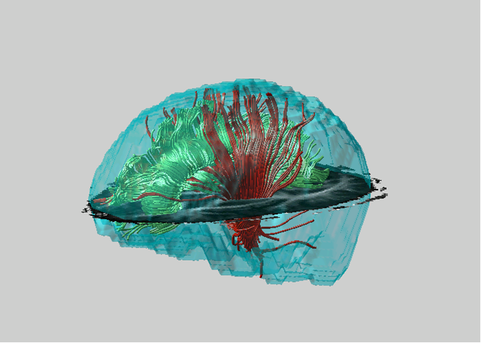
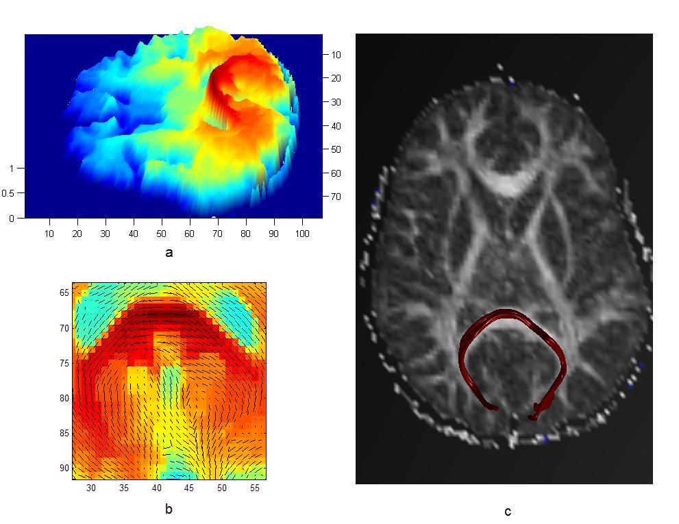
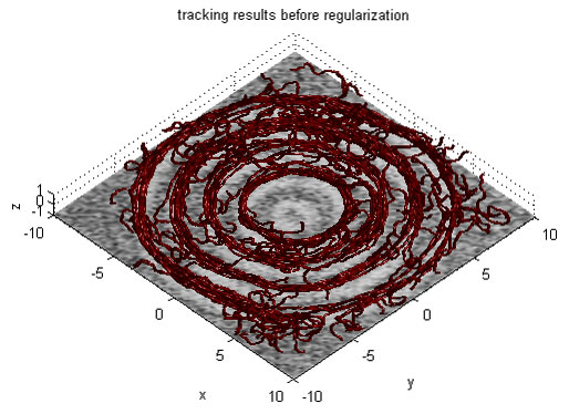
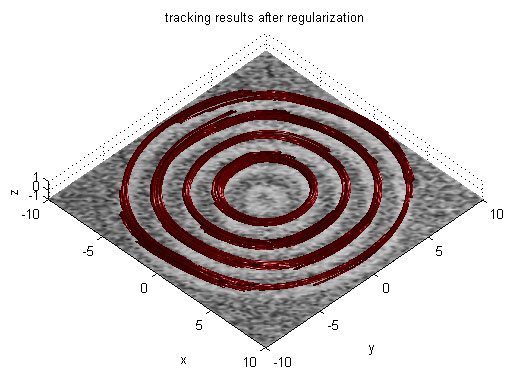
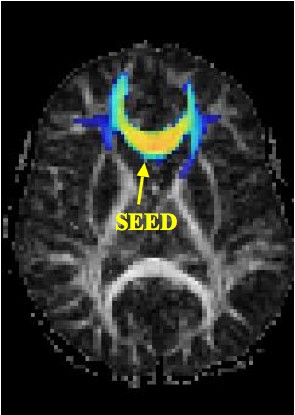
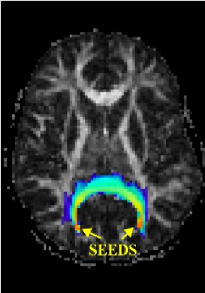
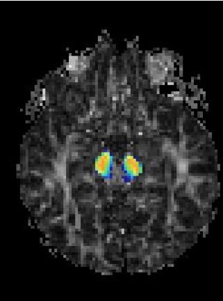
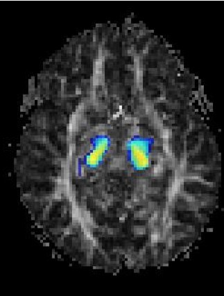
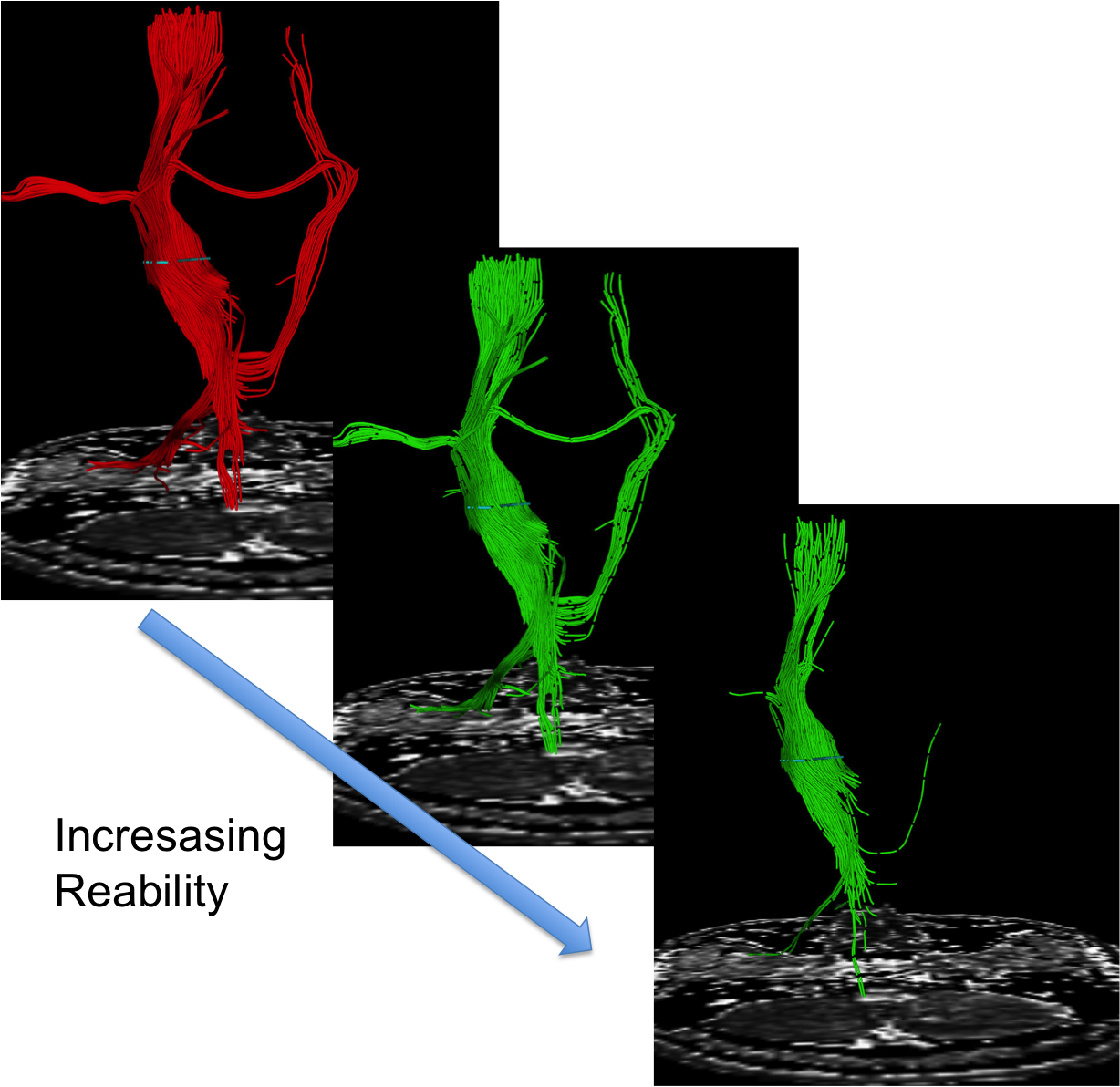
A vector field structure sensitive function, working on the noisy vector field, was proposed that has different responses to 4 types of regions on the data: Non-fiber regions, Outer boundary of the fiber bundles, Inner boundary of the fiber bundles and Fiber regions. As part of the nature of the problem, the outer and the inner boundaries of the fiber bundles are not known exactly (otherwise we would not need to do tracking at all) but rather a region can be marked as a probable outer/inner boundary of the fiber bundles. The regularizing nonlinear diffusion tensors are then built based on this structure sensitive function. The applied nonlinear diffusion is near isotropic in regions far away from the boundaries and anisotropic diffusion (with the principal diffusion direction being parallel to the fiber bundle boundary) in the fiber boundary neighbourhood.
 
This project was partially supported by TUBITAK KARIYER-DRESS (104E035)
LoS: Lattice of Springs Model for Multiple Seed Point Connectivity Maps via Energy Minimization
Erdem Yörük
 
In this work, we propose a model based on a physical setup of nodes (no masses) and springs (whose spring constants were set according to the DTI data). We showed that the connectivity map associated to this physical system can be achieved by minimizing its total potential energy. The variational approach for computing this stationary pattern, has been found to be equivalent to a modified diffusion process. We showed that our method provides connectivity maps that correlate with normal anatomy on real patient data. The proposed method is fast to compute, has a tuning parameter that allows one to adjust the relative importance of the principal diffusion direction among other diffusion directions, allows multiple seed selection to incorporate a-priori information about the anatomy.
 
In general, connectivity can be interpreted as a measure which is proportional to the qualitative similarity and spatial proximity of the units contained in data to be analyzed. In the case of DT images, where we are trying to reveal a functional connectivity, qualitative assessment of tensors becomes crucial in order to construct an appropriate model. We have to consider some certain features embedded in a DT image as indicators of connectivity. In general, the connectivity measure between two tensors can be visualized in terms of the overlap of the distributions they represent. This interpretation will take the relative features of tensors into consideration such as their respective locations, sizes, orientations and shapes. We exploited this idea and used different forms local connectivity that depends on local tensor data. This local connectivity is related to a spring constant and the physical system thus constructed is stabilized (steady state solution) after a perturbation  at seed point(s). The final potential distribution provides the connectivity.
 
Partially supported by TUBITAK KARIYER-DRESS (104E035), by SIMILAR NoE (EU 6th Framework) and by Stanford University, LUCAS MRS/I Center, B.A.P. 05M106.
SMT: Split/Merge Tractography
Ahmet B. Yoldemir, Uğur Bozkaya
 
MR-DTI fiber tractography aims at reconstructing the fiber bundles from MR-DTI data and/or estimating a connectivity map of the brain. The conventional tractography relies on numerical integration of the principal diffusion directions. This approach is prone to accumulating errors, requires careful apriori regularization and does not utilize the complete information embedded in MR-DTI data. Connectivity maps, on the other hand, use the complete MR-DTI information, is more complete but does not provide easily interpretable results, such as fiber (bundles).
 
SMT proposes a new approach to merge the advantages of these two approaches. It is a generic method that is applicable with any tractography output and diffusion model, with appropriate manipulations. The current implementation uses second order tensor modelling of diffusion (DTI) and standard RK4 numerical integartion based tractography. Input tracts are Split and re-Merged, creating a short tract to short tract connectivity matrix, which is later used in interactive visualization based on the reliability of short tract clusters
 
Partially supported by TUBITAK 104E035, TUBA-GEBIP 2008, B.A.P. 07A203 & 10A02R6, Yeditepe University Hospital.
A Pointwise Correspondence Based DT-MRI Fiber Similarity Measure
Sema Berkiten
 
In this work, we proposed a new technique to calculate similarity between fiber tracts which is based on point-wise correspondence. Then we map fiber tracts to R3 with using Laplacian Eigenmaps. The Laplacian Eigenmaps are used to map the tracts into a minimum dimensional embedding space. We have observed a clearly identifiable manifold structure in this R3 embedding space. Consequently, our preliminary results suggest that the point-wise correspondence based fiber tract similarity measure is capable of forming a continuum of fiber tracts (within a bundle) in an implicit embedding space which can be exploited through the use of Laplacian Eigenmaps. The resulting low dimensional manifold can be learned and potentially be used for both outlier fiber tract detection and correction. Further research is required to learn and represent the aforementioned manifold.
 
Partially supported by TUBITAK KARIYER-DRESS (104E035).
 
Figure (left) depicts the manifold structure of the fiber space, embedded in 3D space. Each point coresponds to a fiber, which are colored based on their proximity within the manifold and displayed for corpus collasum. Figure (right) is a pseudo coloring of the corresponding tracts.