 Based on the fact that clinical experience plays a key role in the performance of medical professionals, it is conjectured that a Clinical Experience Sharing (CES) platform, i.e. a searchable collective clinical experience knowledge-base accessible by a large community of medical professionals, would be of great practical value in clinical practice as well as in medical education. Such a CES would be composed of a multi-modal medical case database, would incorporate a Content Based Case Retrieval (CBCR) engine and would be specialized for different domains. Project CaReRa aims at developing such a CES for the domain of liver cases. During the course of the project, multi-modal case data will be collected, anonymized and stored in a structural database, CBCR technologies will be developed, experiments for the assessment of its impact on the clinical workflow as well as medical education will be designed and conducted.
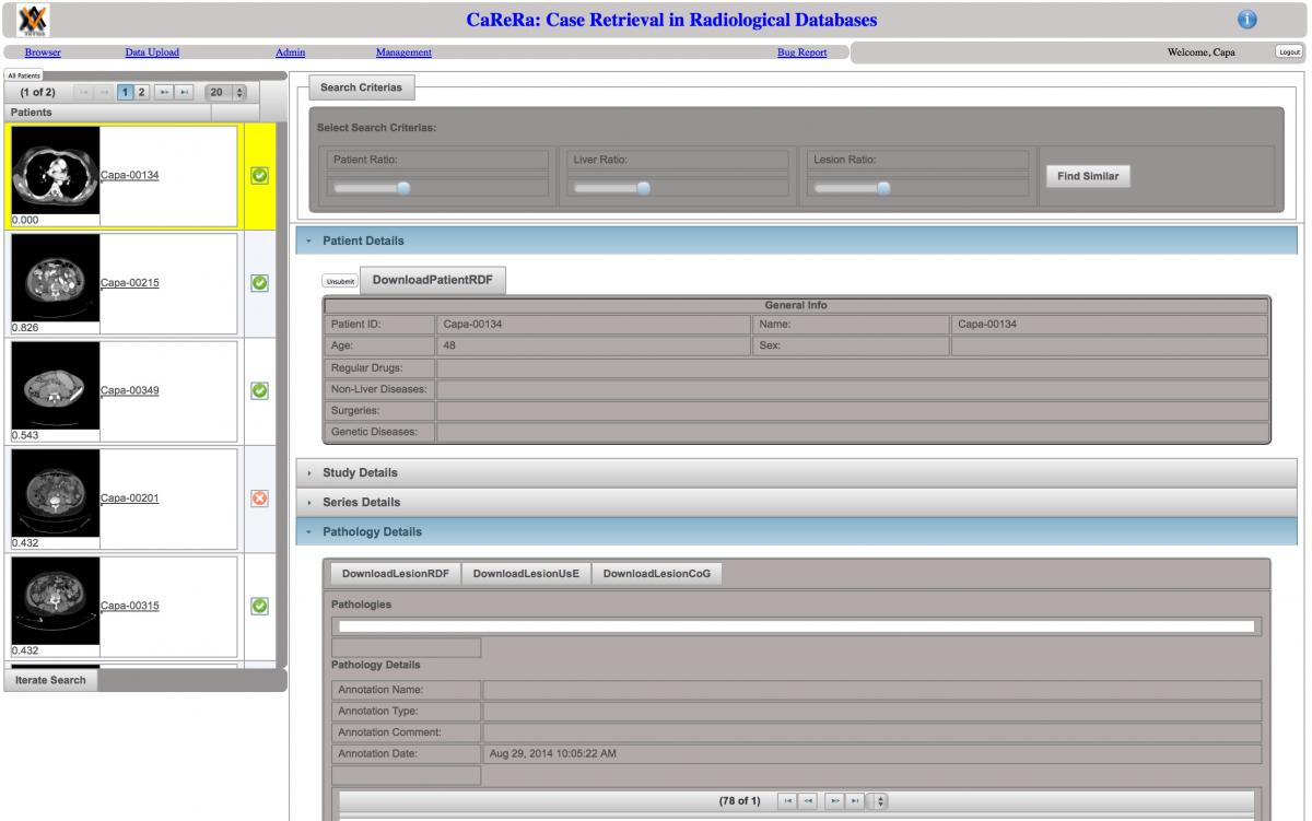
Try our CRR-WEB (if you experience connection problems, please send an email to acarbu@boun.edu.tr) to upload, browse, search and review. Login to "Demo" version, using "demo / demo" as account name and password. IMPORTANT: We suggest you to use Firefox. In your first attempt, your browser may warn you with "Your connection is not secure". In that case, please proceed to "Advanced" options and "Add Exception".
Once you login using demo account, there are two options: Browser and Data Upload. Browser allows you to browse (view) submitted patients. Currently, you can perform conventional search using keywords. The search engine, being developed at CaReRa project, that uses a case as a query will be accesible from this page. Data Upload allows you to upload a new patient to CaReRaWeb. The "Demo" user has limited access and authorization for demonstration of the system.
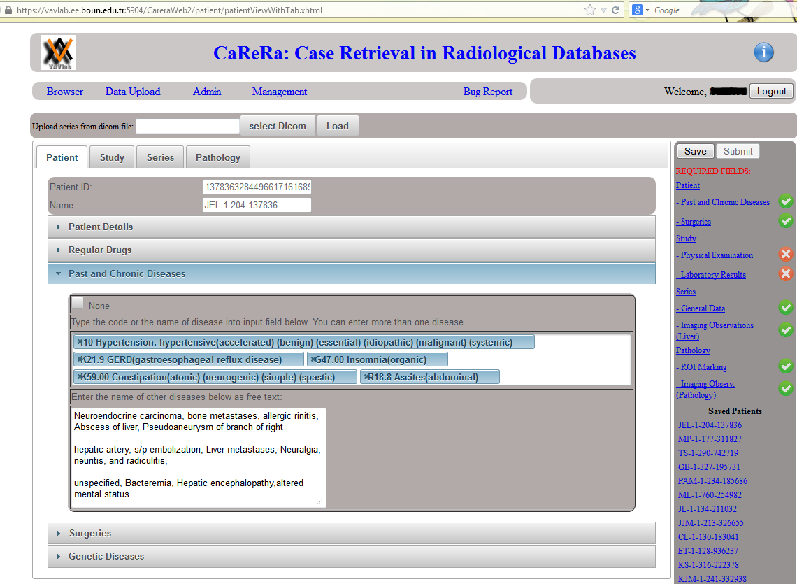
Data Upload: On the right hand side, a list of required fields are displayed. Whenever a required field is filled with some data, a green check mark (otherwise a red cross mark) is shown. Once the radiologist provides information about all required fields, the patient can be submitted. Otherwise, the patient information is saved and can be completed later.
Patient Tab: In this tab, patient information (demographic, regular drugs, past and chronic diseases, surgeries and genetic diseases) is provided. Past and chronic diseases of a patient are listed. Note that diseases are provided using ICD-10 codes. Other diseases are listed as free text.
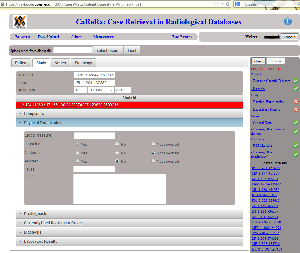
Study Tab: In this tab, patient complaints, physical examination results, prediagnosis, currently used non-regular drugs, diagnosis and laboratory results are provided. Physical examination results are listed. The radiologist's observation is that the patient has "Jaundice" and "Ascites".
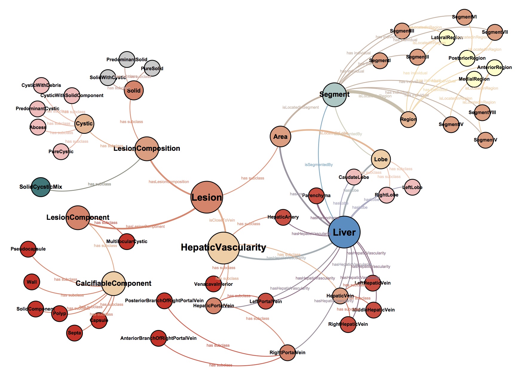
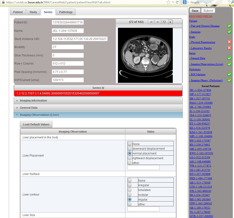
Series Tab: In this tab, imaging observations of liver are described. All the imaging observation fields and values are automatically retrieved from ONLIRA. In this figure, the radiologist reports that the liver position in the body is normal and the liver contour is regular.
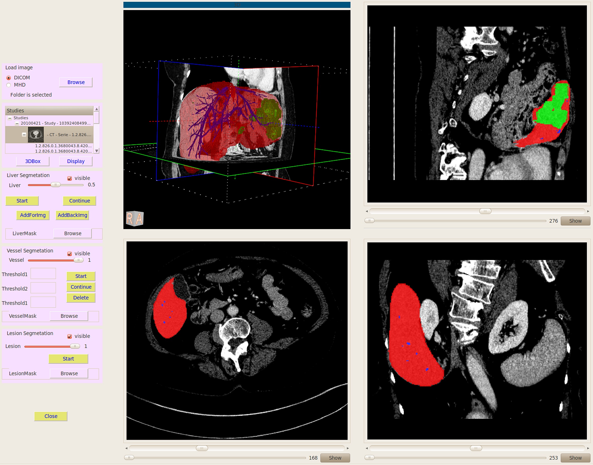
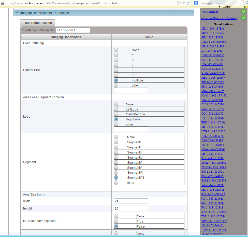
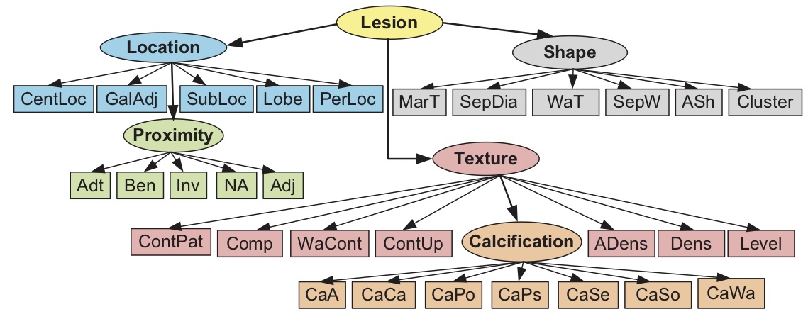
Pathology Tab: In this tab, imaging observations of lesions are described. A DICOM viewer is integrated to CaReRaWeb. The DICOM image is visualized and a rectangular area where the lesion is observed is marked by the radiologist. Then, imaging observations of marked lesion are specified. All the imaging observation fields and values are automatically retrieved from ONLIRA. In this figure, marked rectangular area contains multiple lesions that are located at Segment 8 in the right lobe of the liver. The size of the largest lesion in this area is measured as 21x20mm. This lesion is not adjacent to gallbladder.
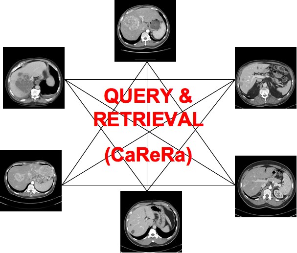
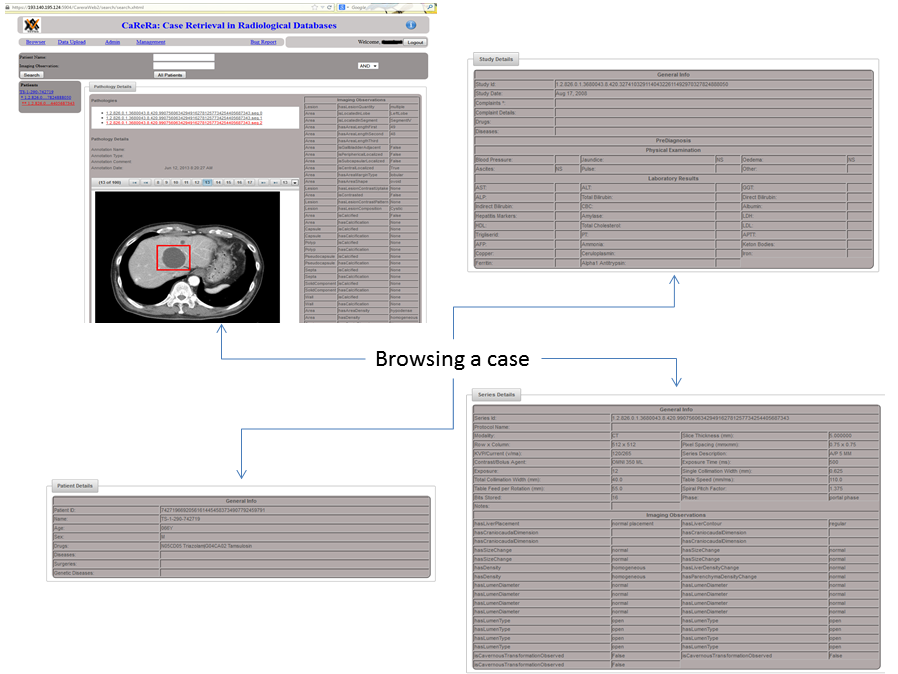
Browser: The radiologist can search in all submitted patients by providing some keywords. Another option is to list all submitted patients.


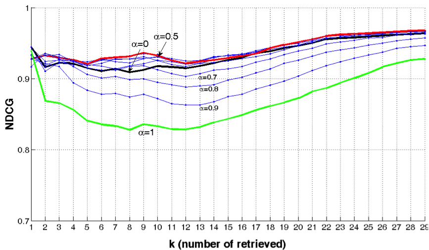
 A radiologist-in-the- loop semi-automatic CMIA system is proposed. It is based on a Bayesian tree structured model, linked to RadLex. The experiments with liver lesions in computed tomography (CT) images. show that on average 7.50 (out of 29) manual annotations is sufficient for 95% accuracy in liver lesion annotations. The proposed system guides the radiologist to input the most critical information in each iteration and uses a network model to update the full annotation online. The results also suggest that the domain-aware models perform better than the domain-blind models learned from data.