BRAINet: Structural and Functional Brain Network Analysis

The idea that the brain is composed of functional and structural networks and that defects of these networks are either the cause or the result of the majority of neurological/psychiatric diseases, is widely accepted and is supported by studies on different patient groups. The use of fMRI (functional MRI) for studying the functional networks (fNETs), and dMRI (diffusion MRI) for studying the structural networks (sNETs) have increased within the last decade. Despite the advantages of these modalities, there are multiple open problems in brain network modeling. Some of the major challenges are:


  • Functionally homogeneous, spatially consistent across a population brain parcellation
  • Directed, weighted and dynamic structural and functional connectivity definitions
  • Spatial resolution limits of dMRI that limits the assessment of brain microstructure in-vivo
  • Temporal resolution limits of fMRI that limits the assessment of brain dynamics
  • Structure-Function relation
  • Machine Learning for graphs
  • Clinical translations for early diagnosis, disease monitoring, pharmacological studies


BRAINet is a series of projects endeavoring to tackle with these problems within a unified functional and structural network modeling setup. We have chosen the Alzheimer’s Disease as our primary application area, though the methods are not disease specific. The project involves researchers from different institutions and fields, including engineering (Electrical-Electronics Eng., Computer Science), basic sciences (Physics, Mathematics) and medicine (Neurology, Physiology). The team uses I.U. Hulusi Behcet Life Sciences Center MRI facilities for data collection.

Watch the short video published by Bogazici Buzz

Related publications

Selected Literature

Diffusion MRI & Tractography:

  1. Le Bihan, D, et al., Artifacts and pitfalls in diffusion MRI, JOURNAL OF MAGNETIC RESONANCE IMAGING (2006)
  2. Basser, PJ, et al, Diffusion-tensor MRI: theory, experimental design and data analysis - a technical review, NMR IN BIOMEDICINE (2002)
  3. Wu, WC, et.al., Image Formation in Diffusion MRI: A Review of Recent Technical Developments, JOURNAL OF MAGNETIC RESONANCE IMAGING (2017)
  4. Alexander, DC, et al., Imaging brain microstructure with diffusion MRI: practicality and applications, NMR IN BIOMEDICINE (2019)
  5. Jeurissen, B., et al., Diffusion MRI fiber tractography of the brain, NMR IN BIOMEDICINE (2019)
  6. Mori, S., et al., Fiber tracking: principles and strategies - a technical review, NMR IN BIOMEDICINE (2002)
  7. Jenkinson, M., et al., FSL, NEUROIMAGE (2012)
  8. Tournier, JD, et.al., MRtrix3: A fast, flexible and open software framework for medical image processing and visualisation, NEUROIMAGE (2019)

Brain Connectomes:

  1. Sporns O, Structure and function of complex brain networks, Dialogues Clin. Neuroscience (2013 Sep 1)
  2. Bullmore E, Sporns O, Complex brain networks: graph theoretical analysis of structural and functional systems, Nat Rev Neurosci (2009 Mar 1) 10: 186-98
  3. Fornito A, et al, Graph analysis of the human connectome: promise, progress, and pitfalls, Neuroimage (2013 Oct 15) 80: 426-44
  4. Kaiser Marcus, A tutorial in connectome analysis: Topological and spatial features of brain networks. NeuroImage (2011 Jan 1) 57: 892-907
  5. Zhang, F., et al., Quantitative mapping of the brain's structural connectivity using diffusion MRI tractography: A review, NEUROIMAGE (2022)
  6. Zalesky A, Fornito A, Bullmore ET. Network-based statistic: identifying differences in brain networks. NeuroImage (2010) 53:1197-207
  7. Hagmann P, et al, Mapping the Structural Core of Human Cerebral Cortex. PLoS Biology (2008 Jan 1) 6: e159
  8. Bastiani M, et al, Human cortical connectome reconstruction from diffusion weighted MRI: The effect of tractography algorithm. Neuroimage (2012 Jun 12) 62: 1732-1749
  9. Smith SM, et al, Network modelling methods for FMRI. Neuroimage (2011 Jan 15) 54: 875-91
  10. Horn A, Blankenburg F. Toward a standardized structural-functional group connectome in MNI space. Neuroimage (2016 Jan 1) 124 (Pt A): 310-322.
  11. Zhu Dajiang, et al, Fusing DTI and FMRI Data: A Survey of Methods and Applications. NeuroImage (2013 Jan 1)
  12. Griffa A, Baumann PS, Thiran JP, Hagmann P. Structural connectomics in brain diseases.  Neuroimage (2013 Oct 15) 80: 515-26
  13. Cichocki A.Tensor Decompositions: A New Concept in Brain Network Analysis? (2013) http://arxiv.org/abs/1305.0395
  14. Zhang Z, Allen GI, Zhu H, Dunson D. Tensor network factorizations: Relationships between brain structural connectomes and traits. Neuroimage (2019 Aug 15) 197: 330-343
  15. Preti M, Van De Ville D. Decoupling of brain function from structure reveals regional behavioral specialization in humans. Nature Communications, (2019), 1-7, 10(1)
Graphs:
  1. Graph Signal Processing, Schuman et al, 2013
  2. Representation Learning on Graphs, Hamilton et al. 2017
  3. Stanford Graph Learning Workshop, 2022
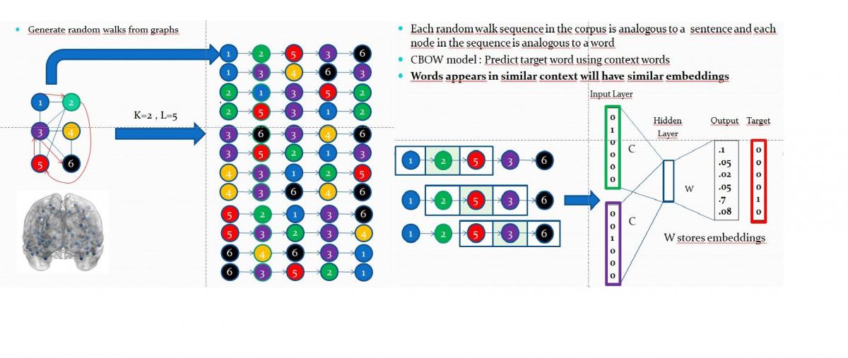
  4. Graph Neural Networks (GNNs)
    • GRAPHSAGE: A fundamental messaging approach
    • GCN: Graph Convolutional Networks
    • ChebyNET: En efficient spectral GCN
    • GIN: Graph Isomorphism Networks
    • GAT: Graph Attention Transformers
Core Collaborators

BRAINet Preprocessing Pipeline Overview


BRAINet projects approach to connectome analysis is based on exploiting multi-modal connectome, as such all modalities need to be co-registered before proceeding to connectome construction and analysis. Co-registration of multi-modal data is a challenging task without any general purpose solution. Hence, we pursue a customized pre-processing pipeline that is designed to utilize the best components of well-known software toolboxes. Primarily, we use FSL for fMRI processing and registration of scalar volumes (such as T1), FreeSurfer for parcellation (atlas registration) and Tortoise for DWI processing. All co-registered volumes are loaded to the BRAINet Platform for manual/visual verificiation by expert neurologists as the final quality check. Cases labeled as poorly co-registered by the neurologists are excluded for further processing and are not manually corrected. Hence, all data used in BRAINet has been identically pre-processed without manual intervention.

BRAINet Platform (An MITK Plug-in)
D. Yüksel, G. Durusoy, A. Karaaslanlı, Z. Kahraman, K.E. Özgün, G. Gümüş
The BRAINet platform is a custom plug-in developed under MITK (www.mitk.org). The platform has been designed to load and display fMRI, DWI, T1, T2, Parcellation, Segmentation and  precomputed ICA-maps, to perform DTI reconstruction, to run deterministic and probabilistic tractography on DTI, to perform network node definition & refinement, to generate multi-connectivity sNET and fNET connectomes.
 
B-Tensor Factorization for Alzheimer's Disease Diagnosis and Understanding
Goktekin Durusoy

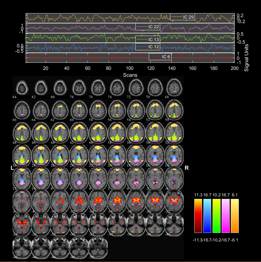
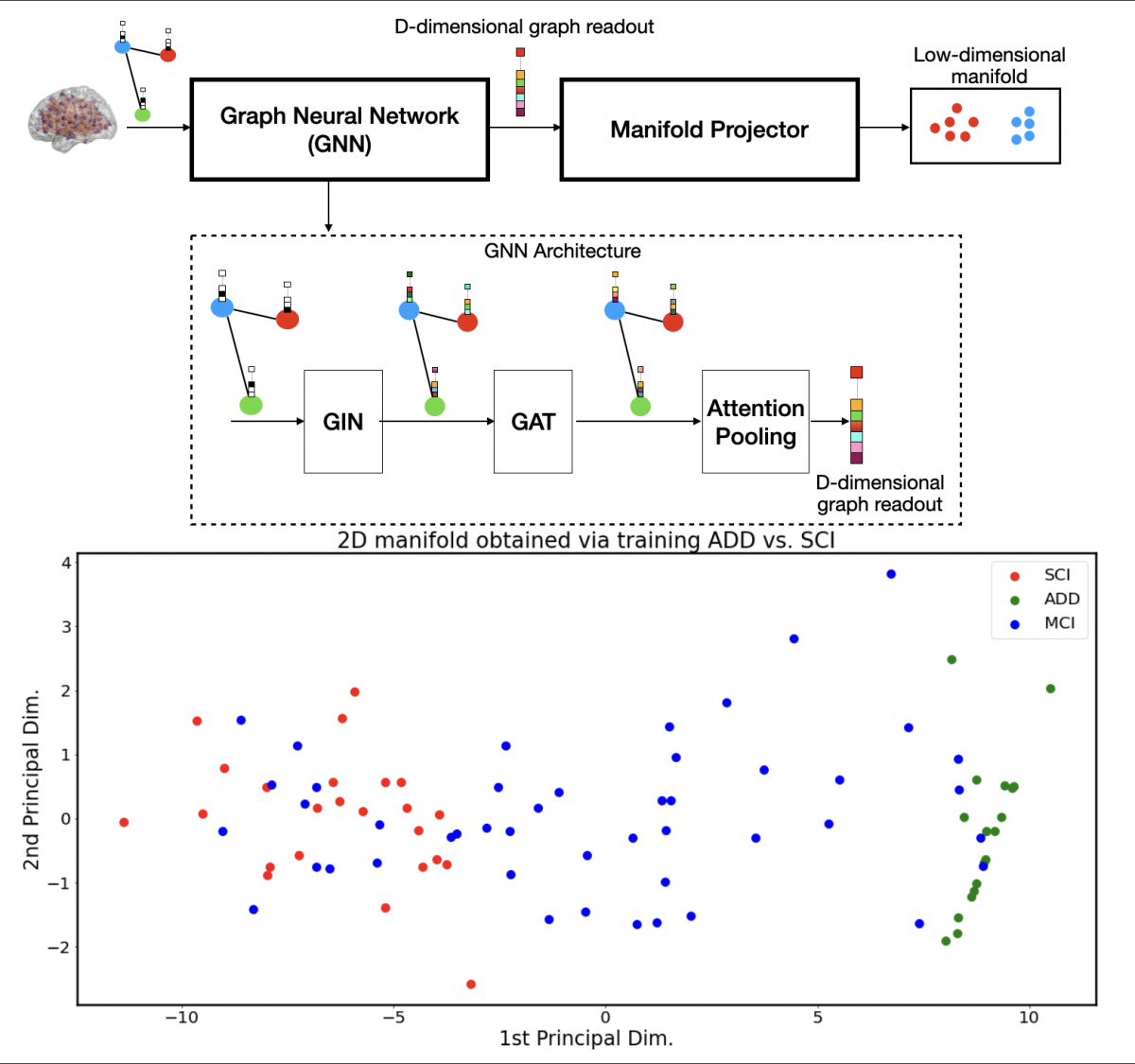
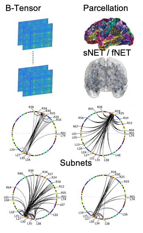
A promising approach in AD research is the analysis of structural and functional brain connectomes, i.e. sNETs and fNETs, respectively. We propose to use tensor representation (B-tensor) of uni-modal and multi-modal brain connectomes to define a low-dimensional space via tensor factorization. The proposed B-tensor encapsulates the whole population either with uni-modal (3D Tensor) or multi-modal (4D Tensor) networks. We show on a cohort of 47 subjects, spanning the spectrum of dementia, that diagnosis with an accuracy of 77% to 100% is achievable in a 5D connectome space using different structural and functional connectome constructions in a uni-modal and multi-modal fashion. We further show that multi-modal tensor factorization improves the results suggesting complementary information in structure and function.

A neurological assessment of the connectivity patterns identified largely agrees with prior knowledge, yet also suggests new associations that may play a role in the disease progress. The precuneus (L/R30), the angular gyrus (L/R25), the supramarginal gyrus (L/R26), the superior parietal lobule (L/R27), the precentral gyrus (L/R29), the postcentral gyrus (L/R28), the dorsal & ventral parts of posterior  cingulate cortex (L/R 9-10) are observed to be the most conspicuous regions.

Assessment of Functional Connectivity Methods in Dementia
Başak Kılıç

Using resting-state fMRI to investigate functional connectivity measures and detect abnormality within and between resting-state networks have yielded promising results that disclose information about the nature of neurodegenerative diseases. The main motivation behind this work was to understand the changes of functional connectivity measures within the components of Default Mode Network (DMN) for people suffering from dementia. The analyses were conducted on three subject groups: subjective cognitive impairment (SCI), mild cognitive impairment (MCI) and Alzheimer’s disease (AD).  By using varying resting-state fMRI methods, such as seed-based, independent component and cluster analyses, it was possible to differentiate between SCI, MCI and AD patients by investigating the dementia related changes within the DMN. Independent of the method of choice, the obtained results indicated a similar pattern of change in connecitivity measures that showed significant differences between each group.


fMRI Guided Personalization of Cortical Parcellation Maps
M.S. Onay, U. Küçükarslan

Joint analysis of structural and functional brain connectomes (sNET & fNET) requires networks to be defined over the same network nodes, ie. cortical parcellation regions. Such an analysis assumes that each network node (cortical parcellation) is spatially well-defined to assure inter-subject correspondence (which is provided by using an atlas) and is functionally compact (ie. high intra-parcel functional connectivity). Here, we propose to use a 3D elastic warping strategy (inspired by the Demon's algorithm) to iteratively modify cortical parcellation maps to increase intra-parcel functional connectivity. Results suggest a statistically significant increase in intra-parcel BOLD signal correlations.
 
Figure: A sample refined parcellation map and intra-parcel mean BOLD signal correlations before and after refinement for healthy individuals and AD patients.
BRAINet Platform Integration with High-Res Schaefer Parcellation Atlases
F. Soğukpınar, A. Hayran


Cortical parcellation is required to define a common node set for consistent joint analysis of structural and functional networks over a population. The built network models are limited by the resolution of the parcellations used. Too coarse parcellations are poor in localization of functional/structural units of the brain, while too high parcellations (voxel-level analysis at the extreme end) prohibit population studies due to difficulties in across-subject correspondences. The Destrieux atlas (148 parcels) has been commonly used. In this work, the recently developed Schaefer atlas (A. Schaefer et al, Cerebral Cortex, 28, 2018) with multi-resolution parcellations (100-1000) has been integrated with BRAINet. Initial comparisons of global network parameters between 148 and 400 node networks showed high correlation, confirming the usability of the new atlas in BRAINer network analysis.


A Preliminary Study of Brain Tensor (B-Tensor) Factorization Reveals Salience Subnetwork as a Strong Discriminator
Göktekin Durusoy

Global brain network parameters suffer from low classification performance and fail to provide an insight into the neurodegenerative diseases. Besides, the variability in connectivity definitions poses a challenge. We propose to represent multi-modal brain networks over a population with a single 4D brain tensor (B) and factorize B to get a lower dimensional representation per case and per modality. We used 7 known functional networks as the canonical network space to get a 7D representation. In a preliminary study over a group of 20 cases, we assessed this representation for classification of Alzheimer's Disease patients from normals. We used 6 different structural connectomes (different connectivity definitions). Linear discriminant analysis results in 90–95% accuracy in binary classification. The assessment of the classification hyperplane revealed Salience subnetwork to be the most powerful in classification consistently over all connectivity definitions. The method was presented at CNI'18 (in conjunction with MICCAI 2018). Our research direction is towards identifying subnetworks as well as fusing structure and function in this framework.
 
Figure shows the classification accuracies for 6 different structural connectomes (the top 2 are weighted and normalized weighted tract count based connectivitiesm while others are based on local diffusivity statistics) when projected onto one or multiple of 7 functional networks (The salience subnetwork -C4- is consistently the most discriminative network)


Nodal Brain Connectome Embeddings
Gurur Gamgam
We propose a random walk based nodal embedding technique for structural brain connectomes in order to represent nodes (cortical regions) in a low dimensional space that reflects their local structural connectivity patterns. Proposed method is inspired by natural language models and treats nodes as words. Thus each connectome (individual) corresponds to a different language with a common vocabulary (node set). Firstly a corpus is formed from random walk sequences (analogous to sentences), then it is fed into a neural probabilistic language model and nodal embeddings (analogous to word embeddings) are obtained. The resulting nodal embeddings are used individually for binary discrimination of Alzheimer's Disease, Mild Cognitive Impariment  and Subjective Cognitive Impairment cases. Mean classification accuracies of best performing 5 nodes are 0.81/0.78/0.70 for AD-SCI/AD-MCI/MCI-SCI. Further, a shift in best performing nodes is observed suggesting a shift of affected cortical regions throughout the spectrum of dementia.